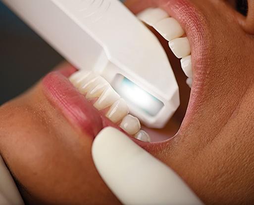
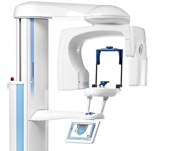
Your doctor can create a 3D image of your teeth in just minutes using an iTero scanner, a leading digital scanner from the makers of Invisalign® clear aligners. The iTero Element Intraoral Scanner is designed to fit your practice even better than before. Whether you are a general practitioner or orthodontist, iTero Element is designed to deliver speed, reliability, intuitive operations, and outstanding visualization capabilities.
Accuracy
Fewer redos and a better aligner fit than with goopy physical impressions.
Invisalign Progress Assessment
Track your progress at every appointment.
Precision
Takes 6,000 images per second.
3D
Get a high-resolution, interactive image of your teeth.
Invisalign Outcome Simulator
See new smile before you even start.
Comfort
No goop. No gagging.
Continuous scanning design lets you scan in motion - eliminating the need to click each time you want to capture a scan. The software automatically detects and repositions scanning start and stop points when you move to a new scanning position within the scanned segment. And while you are scanning, iTero Element is engineered to simultaneously process the scan. It automatically stitches together images for rendering in the correct order, adapts to changes in positioning, and detects and removes soft tissue. Capture everything. View exactly what you need to see.
Scan in colourColour scanning gives you a significant leap forward in visualization. The colour sensor is integrated in iTero Element and the patented dual-aperture lens system is designed to simultaneously capture 2D images in colour with highly accurate 3D laser scanning. Colour scanning can make it easier to distinguish between gingival and tooth structures for a more precise clinical evaluation.
Scan and save dataiTero Element is designed to automatically save scan data to the system's hard disk every two seconds. There’s no need for a battery backup. Even in the event of a power outage, scan data is safe, so you don’t have to worry about losing your work.
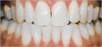
Before
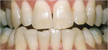
After